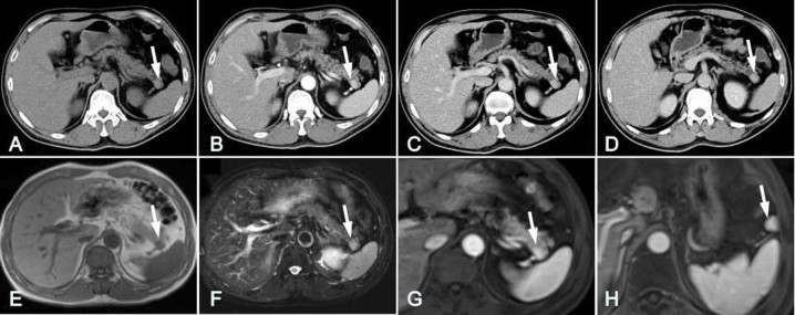
Figure 1.
Case 5. CT and MRI images of a 50-year-old male with IPAS. (A) In the pre-contrast CT image, slight hyper-attenuation of the lesion was visible in the pancreatic tail compared with the pancreas (arrow). Contrast-enhanced axial CT images were obtained in the (B) arterial phase, (C) portal venous phase and (D) delayed phase. High attenuation was shown for the IPAS (arrows) compared with the pancreas in all phases, and the attenuation level of the IPAS was comparable with that of the orthotopic spleen. The lesion (arrow) had (E) a low signal level in T1-weighted imaging and (F) a high signal level in T2-weighted imaging. (G) In gadolinium-enhanced MRI of the arterial phase, the lesion was clearly enhanced. The SI of the nodule was comparable to that of the spleen. (H) In MRI at the splenic hilum level, a triangular accessory spleen (arrow) was visible with a degree of enhancement comparable to that of the orthotopic spleen. CT, computed tomography; MRI, magnetic resonance imaging; IPAS, intrapancreatic accessory spleen.

