. 2018 Jul 3;9(31):6490–6496. doi: 10.1039/c7sc05266a

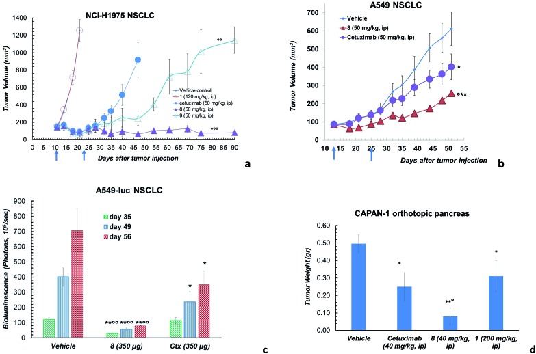
Fig. 3. (a) Effects of ADC 8 and 9 in tumours developed in Nu/Nu mice after s.c. injection of 5 × 10⁶ NCI-H1975 cells. Lesion development and response to antibody treatment was monitored using a Vernier calliper. Mice injected i.p. with either 8, 9, Ctx (4 doses of 50 mg kg–1 once every 4 days) or 1 (4 doses of 120 mg kg–1 once every 4 days) and PBS (n = 8 mice/group; mean and SEM, °°°p < 0.001 and °°p < 0.01 vs. Ctx alone, Mann–Whitney's test). (b) Effects of ADC 8 in tumours developed in Nu/Nu mice for 13 days after s.c. injection of 5 × 10⁶ A549 cells. Lesion development and response to antibody treatment was monitored using a Vernier calliper. Mice injected i.p. with either 8 and Ctx (4 doses of 50 mg kg–1 once every 4 days) or PBS (n = 10 mice/group; mean and SEM, °p < 0.05 vs. Ctx; **p < 0.01 and *p < 0.05 vs. vehicle, Mann–Whitney's test. (c) Artificial metastatic lung cancer experiment carried out with 5 × 10⁶ A549-luc-C8 (A549luc) cells into the tail vein of immunodeficient SCID/beige mice. Tumour bioluminescence imaging (BLI) was recorded by Xenogen IVIS Imaging System 200, at different time points (+35, +49 and +56 days from cell injection), after i.p. injection of luciferin (150 μg per mouse). Mice were treated by aerosol with PBS or ADC 8 or Ctx (3.5 mL of 100 μg mL–1 solution) q7dx4 (n = 12 mice/group; mean and SEM, °°p < 0.01 vs. cetuximab; *p < 0.05 and **p < 0.01 vs. vehicle). (d) Orthotopic tumour pancreas experiment performed with 1 × 10⁶ tumor Capan-1 cells injected into pancreas. Tumor weight was evaluated 90 days after tumor injection. Mice treated intraperitoneally with 8 or Ctx (4 doses of 40 mg kg–1 once every 4 days), PBS and 1 (200 mg kg–1, q4dx4) (n = 10 mice/group); mean and SEM, °p < 0.05 vs. Ctx,*p < 0.05 and **p < 0.01 vs. vehicle).

Fig. 3