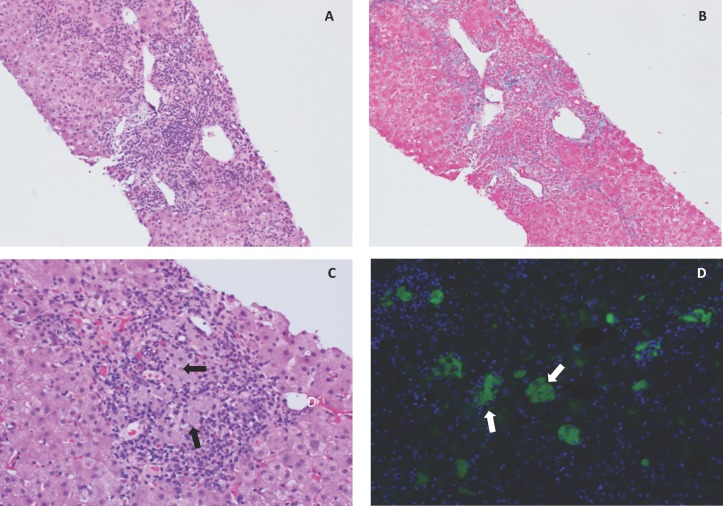
Figure 2.
Photomicrographs of H&E (A; 10×) and trichrome (B; 10×) stained core liver biopsies, demonstrating areas of moderate to marked acute and chronic portal hepatitis associated with piecemeal necrosis and mild portal fibrosis with focal sparse bridging. Within areas of portal inflammation were focal areas of admixed grey-pigment containing histiocytes (C, arrows; H&E, 20×), which was autofluorescent (D; green autofluorescence (excitation 470 nm; emission 475–550 nm), with overlay of blue 4′,6-diamidino-2-phenylindole (DAPI) nuclear counterstain).

