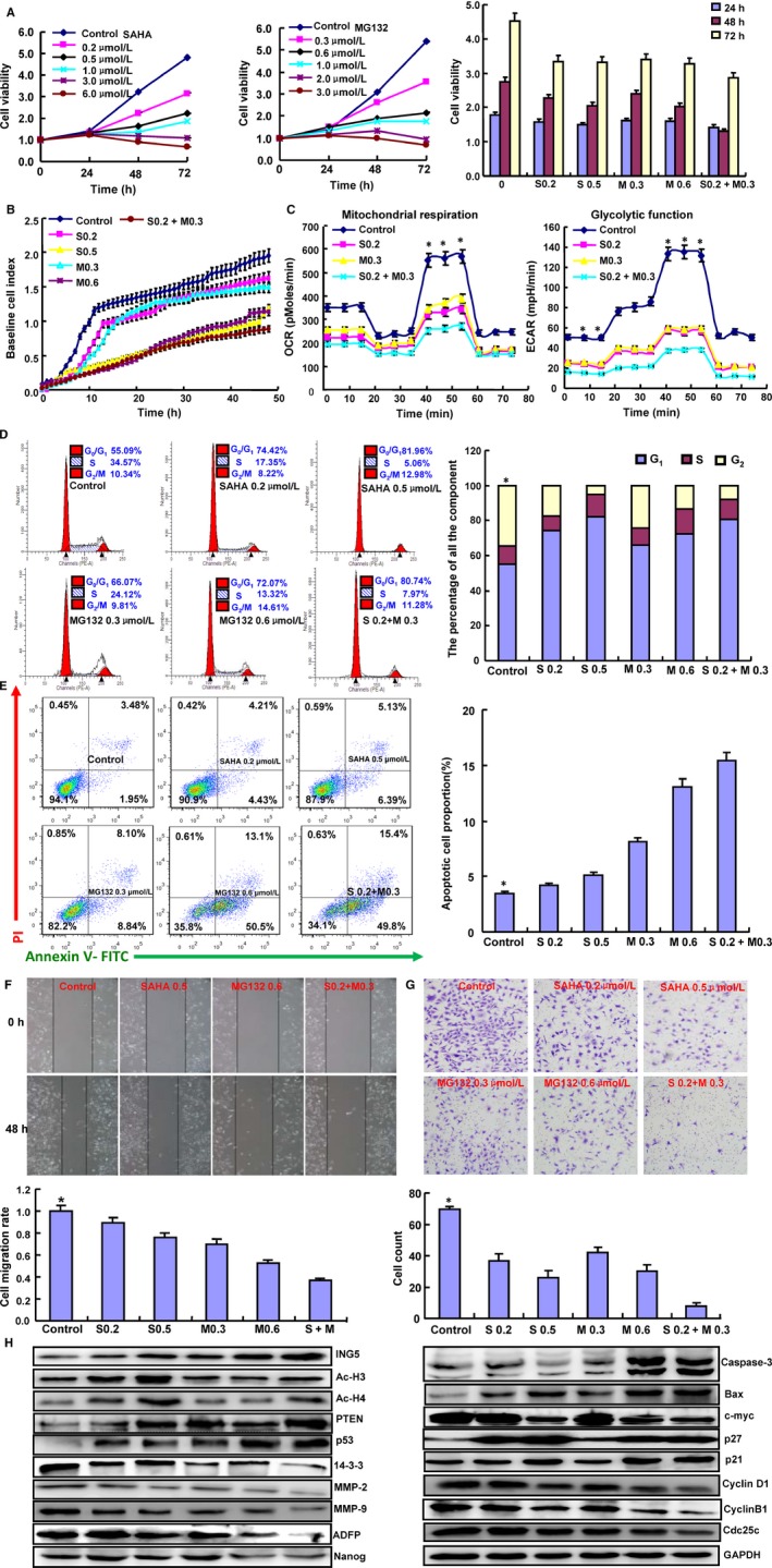
Figure 1.

Effects of SAHA and MG132 on the biological events of SH‐SY5Y cells. Cell proliferation was determined by CCK‐8 (A) and xCELLigence assays (B). The cellular energy metabolism assay detected the rate of oxygen consumption (OCR) and extracellular acidification (C). Flow cytometric analysis was used to detect the cell cycle (D) and apoptotic induction (E). Wound healing (F) and Transwell assays (G) were used to detect the migration and invasion abilities of SH‐SY5Y cells. The protein expression levels of phenotypes were screened by Western blot (H). Note: S0.2, SAHA 0.2 μmol/L; M0.3, MG132 0.3 μmol/L; S0.5, SAHA 0.5 μmol/L; M0.6, MG132 0.6 μmol/L; S0.2+ M0.3, SAHA 0.2 μmol/L and MG132 0.3 μmol/L. *P < .05 vs the treatment group. Each bar represents the mean ± standard deviation of three experiments, with the control as “1.” *P < .05 vs SAHA/MG132 treatment
