Figure 3.

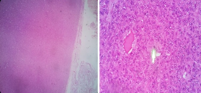
H&E stain of the left parietal mass showing a well-defined, eosinophilic staining tissue with interspersed blood vessels. Green arrow pointing to an area of a mitotically active nucleus.

H&E stain of the left parietal mass showing a well-defined, eosinophilic staining tissue with interspersed blood vessels. Green arrow pointing to an area of a mitotically active nucleus.