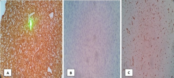
Figure 4.

Immunohistochemistry staining of the left parietal mass revealed the following: (A) smooth muscle antigen—positive tumour cells (B) epithelial membrane antigen—negative tumour cells and (C) CD34 stain—negative tumour cells. Green arrow pointing to an area of tumor’s vascular supply.
