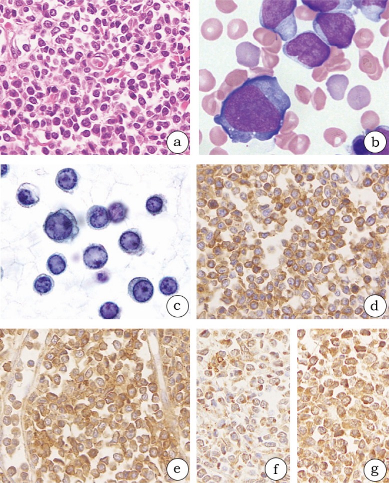
Fig. 1.
Histological and cytological findings in blastic plasmacytoid dendritic neoplasms (1a-g, case 3). (1a) The oval to spindle neoplastic cells diffusely proliferate in the dermis. (1b) The neoplastic cells have scant to modest pale-blue agranular cytoplasm and extend cytoplasmic pseudopodial projections on bone marrow smears. (1c) Neoplastic cells in the pleural effusion specimen have ground glass chromatin, a round nucleus, and some small nucleoli. (1d-g) The neoplastic cells in formalin-fixed and paraffin-embedded specimens from skin lesions are diffusely positive for CD123 (1d), CD303 (polyclonal) (1e), thymic stromal lymphopoietin (1f), and thymic stromal lymphopoietin receptor (1g).

