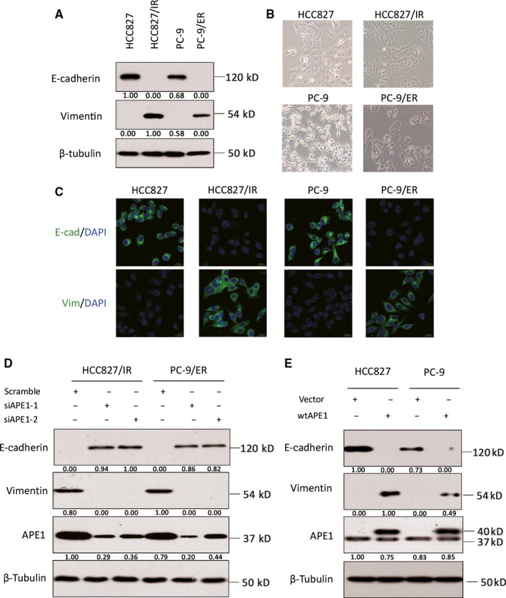
Figure 4.

Manipulations of APE1 regulate the EMT process. The EMT markers in both cell lines with different APE1 status were measured by the epithelial marker E‐cadherin or the mesenchymal marker vimentin using Western blot (A), phase contrast microscopy (B), and immunofluorescent staining (C). Following knockdown of APE1 in HCC827/IR and PC‐9/ER cells via siRNA transfection, the expression of E‐cadherin, vimentin, and APE1 was determined by Western blot (D). Following overexpression of APE1 in EGFR‐TKI‐sensitive cells via lentiviral particles, relative expression levels of E‐cadherin, vimentin, and APE1 were determined by Western blot (E). Representative images or blots from at least three individual experimental repeats are shown in this figure
