Figure 5.

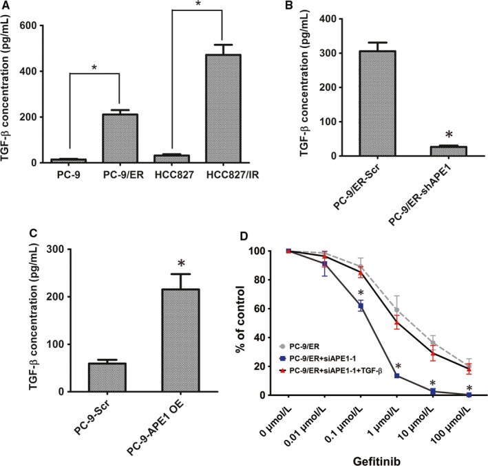
APE1 regulates EMT through TGF‐β signaling. TGF‐β secretion level was evaluated by ELISA in EGFR‐TKI‐resistant cell lines and their parental sensitive cells (A). The 24‐hour cell‐free supernatants of tissue culture from PC‐9 with or without APE1 overexpression and PC‐9/ER with or without APE1 knockdown were collected 48 h post‐transfection or infection and then evaluated by ELISA (B and C). Recombinant TGF‐β protein was added to the culture medium of APE1 knockdown PC‐9/ER cells, treated with increasing concentrations of gefitinib, and then, the cytotoxicity of EGFR‐TKI was determined by CCK8 assay (D). To exclude the impact of APE1 manipulation on cell growth, various gefitinib dose treatments of each group have been normalized to the readout of 0 μmol/L (DMSO only) treatment. Mean values of at least three individual experimental repeats are shown as the mean ± SD. * indicates a statistically significant difference when compared with the same treatment dose as its parental cell (P < 0.01)
