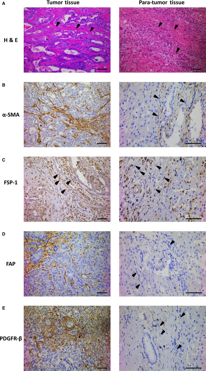
Figure 2.

Expression of α‐SMA, FSP‐1, FAP, and PDGFR‐β in human tumor tissue of ICC and para‐tumor tissue. A‐E, Representative immunohistochemical images of human tumor tissue and para‐tumor tissue stained with H&E (A), α‐SMA (B), FSP‐1 (C), FAP (D), and PDGFR‐β (E) antibodies. CAFs around cancer cells were positive stained for α‐SMA, FSP‐1, FAP, and PDGFR‐β in tumor tissue of ICC, while NFs were negative stained in nontumorigenic tissue (arrowheads; scale bar: 100 μm)
