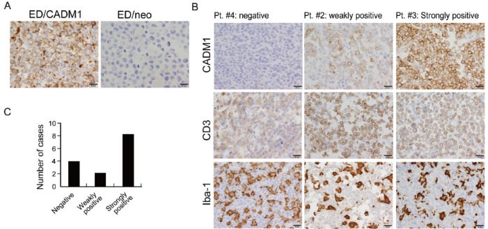
Fig. 3.
Immunohistochemistry of CADM1 in lymphoma cases. (A) The specificity of the anti-CADM1 antibody was tested by using ED/CADM1 and ED/neo cell lines. (B) CADM1 expression in 3 representative cases of ATLL is shown. Lymphoma cells were positive for CD3, and infiltrating macrophages were labelled with anti-Iba-1 antibody. (C) A bar graph shows the total number of cases that were negative, weakly positive or strongly positive for CADM1 expression.

