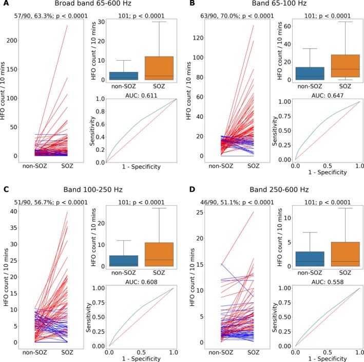
Figure 5.

Pathological high frequency oscillations (pathHFO) are increased in seizure onset zone (SOZ). (A) Broad band pathHFO (65–600 Hz). (B) High gamma band pathHFO (65–100 Hz). (C) Ripple band pathHFO (>100–250 Hz). (D) Fast Ripple band pathHFO (>250–600 Hz). Left Panels (A–D): pathHFO rates (#counts/min‐channel) are increased in SOZ versus non‐SOZ for 63.3% (57/90, sign test P < 0.0001) of epilepsy patients for broad band, 70% (63/90, P < 0.0001) of epilepsy patients in high gamma, 56.7% (51/90, P < 0.0001) of epilepsy patients in Ripple, and 51.1% (46/90, P < 0.0001) of epilepsy patients in Fast ripple band. The lines represent mean HFO rate in non‐SOZ and SOZ of individual patients where (red stands for P < 0.05, sign test). Upper Right Panels: pathHFO rates are increased in SOZ compared to non‐SOZ (P < 0.0001) for all pathHFO bands when electrodes from all epilepsy and facial pain patients are aggregated (N = 102 patients, total electrodes = 6368, SOZ electrodes = 908, nonSOZ electrodes = 5460). Lower Right Panels: Receiver‐operator‐curve (ROC) and area‐under‐curve (AUC) for prospective individual SOZ versus non‐SOZ electrode classification using the pathHFO band rates. The classification of individual electrodes as SOZ using pathHFO rates remains challenging, but most robust in the high gamma band. Note: the support vector machine (SVM) electrode classification using all four pathHFO bands combined with pathHFO amplitude yields a superior electrode classification to any of the individual pathHFO band results.
