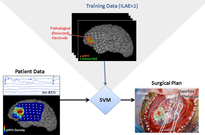
Figure 7.

Support vector machine (SVM) classification of epileptogenic tissue. training data from 31 patients include multiple normalized pathHFO features for each electrode (pathHFO rate, duration, amplitude). Here, SVM uses interictal iEEG data from excellent outcome patients (Engel I) for training. The SVM method uses the array of pathHFO features to train the classifier and then classifies each electrode as pathological (epileptogenic) or normal in an independent testing set (N = 31). In this example, the region of EZ determined by SVM extended beyond the SOZ determined iEEG recording of habitual seizures.
