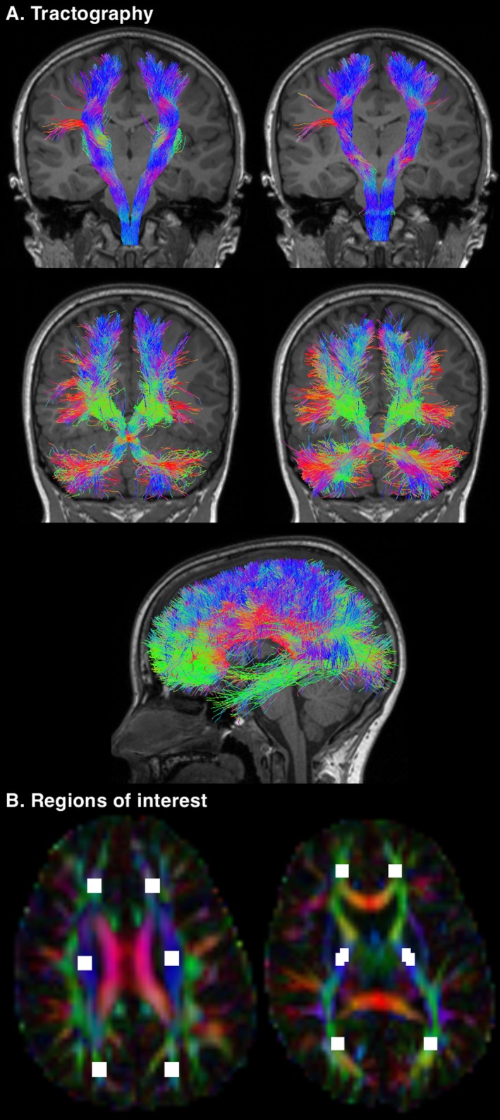
Figure 1.

Illustration of some of the data generated for this study. (A) Tractography performed using the 11‐year diffusion images. Top row: the pyramidal sensorimotor tracts. The corticospinal tract (left) was reconstructed using seed regions in the pons and inclusion regions in the posterior limb of the internal capsule. The somatosensory tract (right) was reconstructed using seed regions in the medial lemniscus, inclusion regions in the thalamus and the combined pericentral cortices (precentral, paracentral, and postcentral cortices), and exclusion regions in the corticospinal tract portion of the pons. Middle row: the cerebellar motor tracts. The cerebellar‐thalamo‐cortical tract (left) was reconstructed using seed regions in the dentate nucleus and inclusion regions in the decussation of the superior cerebellar peduncle, and the contralateral red nucleus, thalamus, and prefrontal cortex. The cortico‐ponto‐cerebellar tract (right) was reconstructed using seed regions in the middle cerebellar peduncle, inclusion regions in the contralateral cerebral peduncle, anterior limb of the internal capsule and prefrontal cortex, and exclusion regions in the decussation of the superior cerebellar peduncle and medial lemniscus. Bottom row: the corpus callosum tracts, reconstructed using automatically generated17, 18 seed regions in the corpus callosum, with the addition of exclusion regions at the brainstem, cerebral peduncle and thalamus. (B) Regions of interest drawn on the 11‐year diffusion images. Six regions were placed on a single superior brain slice at the level of the centrum semiovale (left) and six regions were placed on a single inferior brain slice at the level of the mid‐thalamus (right). These regions of interest were intended to reproduce the regions of interest drawn on the neonatal diffusion images, to enable a comparison of white matter microstructure between the time points. The neonatal regions of interest are shown in Figure 1 of our previous publication.7
