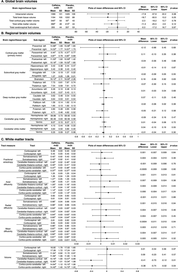
Figure 3.

Global and regional brain volumes and white matter microstructure, contrasted between treatment groups at 11 years of age. Mean differences and P‐values are from separate linear regression models for each MRI outcome, adjusted for age and sex of the child. The plots are visual representations of the mean differences and 95% confidence intervals (CI). For bilateral outcomes, data from the left and right hemispheres were analyzed in a single regression model, and results are presented as a single estimate for the difference for the two hemispheres, as there was little evidence of group by hemisphere interactions. Units are cm3 for volumes and ×10−3 mm2/sec for diffusivities. SD = standard deviation. The analyses are based on the total n = 117 (63 caffeine, 54 placebo) children, although some children are missing from some analyses because their images had artifact which affected that particular analysis: a n = 3 missing; b n = 2 missing; c n = 1 missing; d n = 5 missing; e n = 4 missing. The total numbers are lower for the tractography analyses (n = 116; 62 caffeine, 54 placebo) because one child in the caffeine group did not have b = 3000 sec/mm2 diffusion images acquired and hence had to be excluded from the tractography analyses.
