Figure 5.

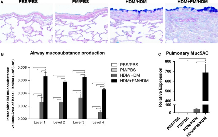
Mucosubstance distribution, abundance, and gene expression. PM exposure during allergen sensitization (HDM + PM/HDM) leads to enhanced mucosubstance secretion upon allergen challenge, compared to allergen‐only sensitized animals (HDM/HDM). (A) Micrographs of paraffin‐embedded lung tissue sections (×400 magnification) stained with Alcian blue and periodic acid‐Schiff (ABPAS). Mucosubstances are stained blue. The scale bar represents a distance of 50 μm. (B) Mucosubstance production was quantified via ImageJ, at four different levels in the lung: Levels 1–4. Mucosubstance production is expressed as intraepithelial mucosubstance volume over the area of basal lamina (μm3/μm2). (C) Pulmonary Muc5ac gene expression was assessed via qPCR. Muc5ac gene expression is shown as relative expression to Gapdh housekeeping gene. Data are presented as mean ± SEM (n = 4/group). Bars indicate a significant difference of P < 0.05.
