Figure 1.
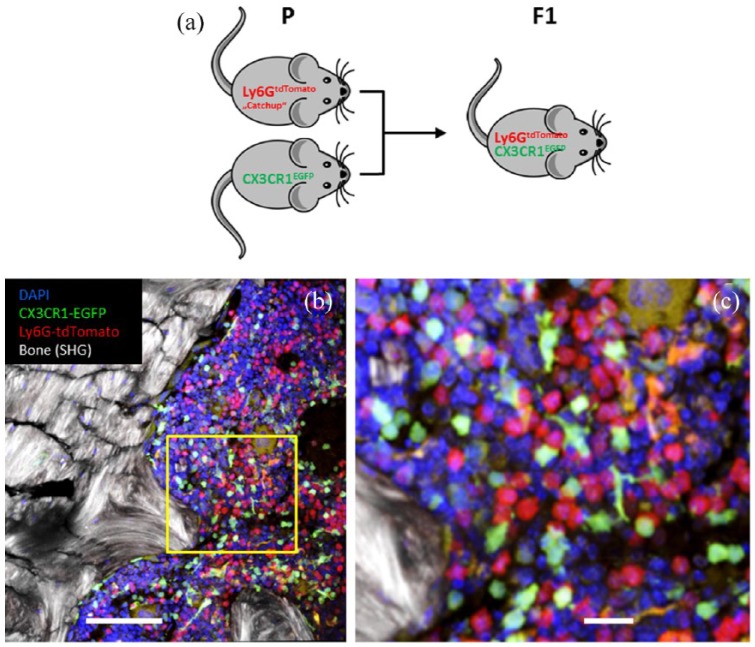
CX3CR1EGFP mice with microglia-specific EGFP expression crossed with Catchup mice show selective expression of tdTomato in polymorphonuclear neutrophils and EGFP staining in macrophages, dendritic cells and microglia.
(a) Breeding scheme for generating mice with red fluorescent PMNs and green fluorescent macrophages/microglia; (b) two-photon microscopy analysis of a section through the bone marrow; and (c) enlarged section in the yellow square. Note, there is no overlap of red (tdTomato) and green (EGFP) staining. The morphology of some green cells is dendritic like, while others are circular. Red cells are uniformly circular. Orange areas are nonspecific autofluorescent structures (not cells). Sections were counterstained with DAPI. Scale bars: 100 µm (b) and 20 µm (c).
DAPI, 4′,6-diamidino-2-phenylindole; F1, first filial generation; EGFP, enhanced green fluorescent protein; P, parental generation; PMN, polymorphonuclear neutrophil; SHG, second harmonic generation signal; tdTomato, red fluorescent reporter protein.

