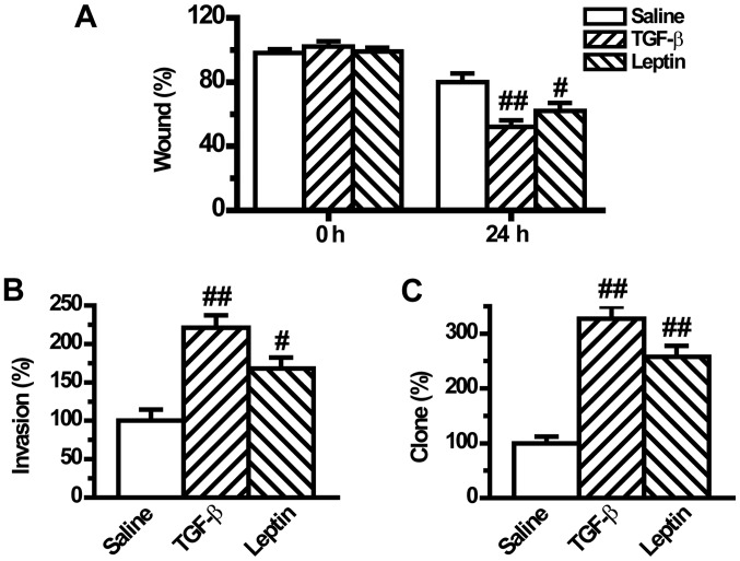
Figure 5.
Leptin enhances epithelial-to-mesenchymal transition-induced tumor phenotypes in A549 cells (n=6). (A) Cell migration was measured by a wound-healing assay. The cells were incubated for 24 h after cell wounding and then the images were captured. (B) The invasive activity of cells was assayed by a Matrigel-coated Transwell assay. The cells that invaded through the filter were quantified 24 h following plating. Data were expressed relative to the invasive ability of the control cells. (C) The tumorigenic phenotype was measured by colony formation assay. All the plotted values are relative to A549 cells in the control group. Values are expressed as mean ± standard error of the mean. #P<0.05, ##P<0.01 vs. the control group.

