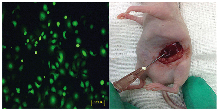
Figure 1.
Establishing a mouse model of liver metastasis from gastric cancer. Representative fluorescence image of MKN45-GFP and representative image of spleen during injection of 1×10⁶/0.2 ml of MKN45-GFP cells into the spleen parenchyma of nude mice. MKN45-GFP cells, stable green fluorescent protein (GFP)-expressing MKN45 cells (scale bar, 50 µm).

