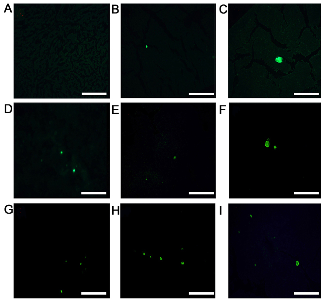
Figure 2.
Representative fluorescence images of low-dose, high-dose and control groups (scale bar, 20 µm). MKN45-green fluorescent protein cells were injected into the spleen parenchyma and the livers were cut into 5-µm thick serial sections for fluorescence imaging. Female BALB/c nu/nu mice aged 4–6 weeks old were assigned randomly into a low-dose group (0.2 µg rmIL-15), high-dose group (2.5 µg rmIL-15) and control group (PBS), with 6 mice/group. All mice were treated five times/week for 3 weeks. On day 28, 6 mice/group were sacrificed. (A-C) Representative fluorescence images of the high-dose group, (D-F) Representative fluorescence images of the low dose group. (G-I) Representative fluorescence images of the control group. rmIL-15, recombinant mouse interleukin-15.

