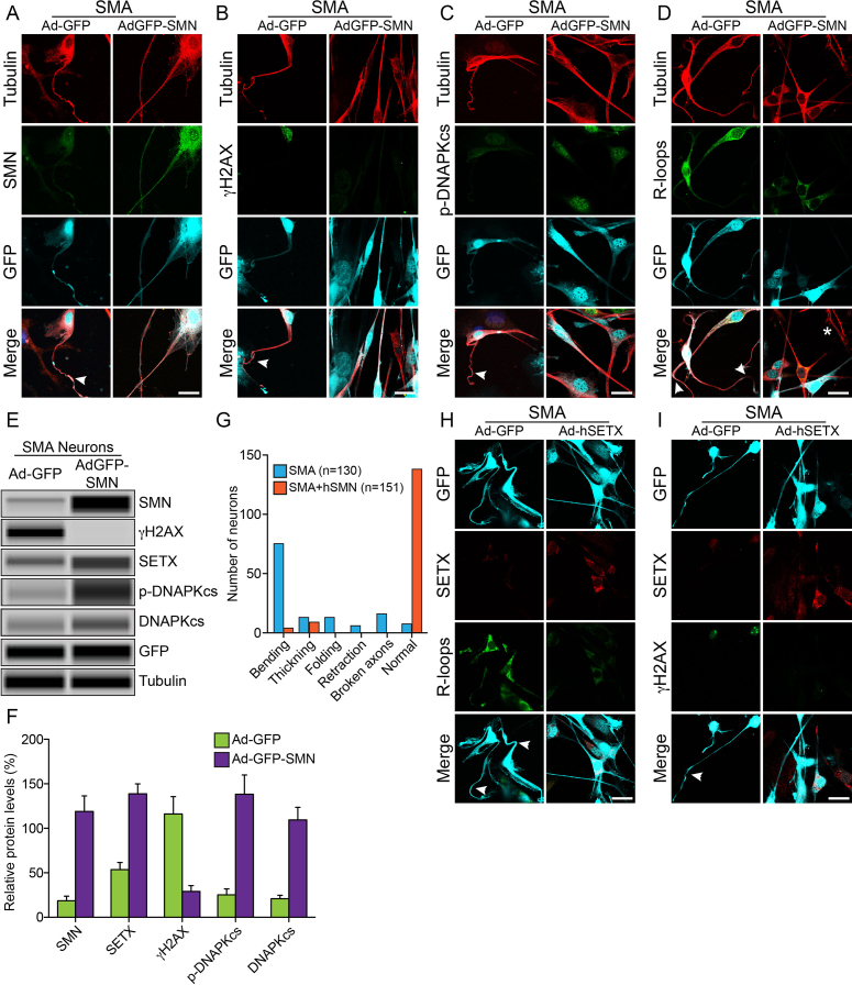
Figure 7.
Rescue of DNA damage and neuron degeneration of SMA spinal cord neurons by ectopic expression of SMN and SETX. Ectopic expression of recombinant SMN restores SETX and DNA-PKcs levels, reduces DDR activation and rescues SMA neuron degeneration. Cultured primary spinal cord neurons were infected adenovirus Ad-GFP and Ad-GFP-SMN1 or Ad-GFP and Ad-h-SETX at 100 MOI for 48 h, fixed with 4% PFA and stained with antibodies and IF examined by confocal microscopy. Low levels of SMN in SMA neurons causes axonal defects, bending, folding, retraction and ballooning (arrowheads), as shown by staining with neuron-specific β-tubulin-III, (clone TUJ1) (red) that lead to degeneration of SMA motor neurons. GFP positive neurons are pseudo-colored as cyan (GFP). Staining for markers of DDR and DNA repair pathways shows restoration of SMN levels, increased levels of DNA-PKcs, rescue of DNA damage and axonal degeneration in SMA neurons. (A) β-tubulin-III (red) and SMN (green), (B) β-tubulin-III (red) and phospho-H2AX (γH2AX) (green), (C) β-tubulin-III (red) and p-DNAPKcs (green) and (D) β-tubulin-III (red) and R-loops (green) show reduced staining for γH2AX and increased staining for p-DNA-PKcs, reduced staining for R-loops and rescue of axonal growth defects. (E) Neurons infected with adenovirus Ad-GFP and Ad-GFP-SMN1 were also harvested at 48 h post-infection to make total cell lysate for protein analysis using automated Wes System (ProteinSimple). Representative capillary-blot images of proteins and phospho-proteins are shown (full-length blots are included inSupplementary Figure S14). Tubulin and GFP blots are shown as loading controls. IB analysis shows restoration of SMN levels in SMA neurons (Ad-GFP-SMN), increased SETX, phospho-DNA-PKcs and total DNA-PKcs levels and reduction of phospho-H2AX (γH2AX) levels compared to control SMA neurons (Ad-GFP). (F) Quantitation and comparison of SMN, γH2AX, SETX, p-DNA-PKcs and DNA-PKcs levels in SMA+Ad-GFP and SMA+AdGFP-SMN neurons show rescue of DNA damage in SMN complemented neurons (SMA+ AdGFP-SMN) by marked reduction in γH2AX levels to (29.08 ± 6.44%, P = 0.013) compared to control SMA neurons (116.1 ± 19.53%) that is supported by increase in levels of SETX (85.19 ± 13.76%, P = 0.003), p-DNA-PKcs (113.0 ± 22.55%, P = 0.007) and total DNA-PKcs (88.55 ± 14.68%, P = 0.003) in (SMA+hSMN) neurons compared to Control (SMA+Ad-GFP) neurons. (G) Analysis of morphological features that represent neurodegeneration such as bending (that includes swelling and ballooning), thickening, folding and retraction of axons show reduction in neuron degeneration and improvement in axonal growth after rescue of DNA damage phenotype in neurons. Neurons stained for (H), SETX (red) and R-loops (green) and (I) SETX (red), phospho-H2AX (γH2AX) (green) show reduced staining for γH2AX and R-loops and rescue of axonal growth defects. Scale bar is 20 μm.

