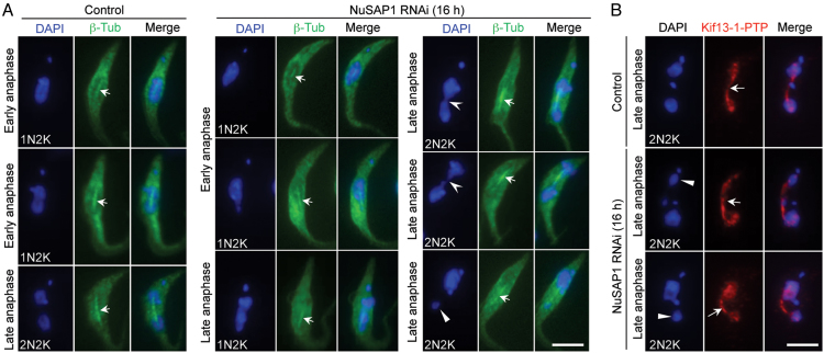
Figure 6.
Spindle formation is not affected in NuSAP1 RNAi cells. (A) Spindle structure in control and NuSAP1 RNAi cells visualized by immunostaining of 3HA-tagged β-tubulin. The solid arrowhead indicates the small nucleus, the open arrowheads indicate the chromosome bridge connecting the two segregating nuclei, and the arrows show the anaphase spindle. Scale bar: 5 μm. (B) Spindle structure in control and NuSAP1 RNAi cells visualized by immunostaining of PTP-tagged Kif13-1. The solid arrowheads indicate the small nucleus, whereas the arrows indicate the anaphase spindle. Scale bar: 5 μm.

