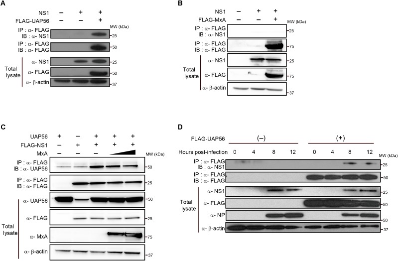
FIGURE 1.
In vitro coprecipitation of NS1 with UAP56 or MxA. (A) In vitro interaction of NS1 with UAP56. HEK293T cells were transfected with protein expression plasmids for WSN-NS1 and/or FLAG-tagged UAP56. At 48 h post-transfection, cells were lyzed and immunoprecipitated (IP) with anti-FLAG antibody. Co-precipitated proteins were analyzed by immunoblotting (IB) with anti-NS1 antibody. (B) In vitro interaction of NS1 with MxA. HEK293T cells were transfected with protein expression plasmids for WSN-NS1 and/or FLAG-tagged MxA. Co-immunoprecipitation and IB were carried out as described in A. (C) Overexpression of MxA does not affect the UAP56–NS1 interaction. HEK293T cells were transfected with plasmids expressing UAP56 and/or FLAG-tagged NS1, and increasing amounts of MxA. Forty-eight hours later, the cells were lyzed and IP with anti-FLAG antibody. Co-immunoprecipitation and IB were carried out as described in A. (D) UAP56–NS1 interaction in influenza A virus-infected cells. HEK293T cells were transfected with pCAGGS-FLAG-UAP56 or a control vector, and infected with WSN virus at a multiplicity of infection (MOI) of one. The cells were harvested and lyzed at the indicated time points post-infection and IP with anti-FLAG antibody. Co-precipitated proteins were analyzed by IB with anti-NS1 antibody; the NP expression levels in the total cell lysate were analyzed as an infection control.

