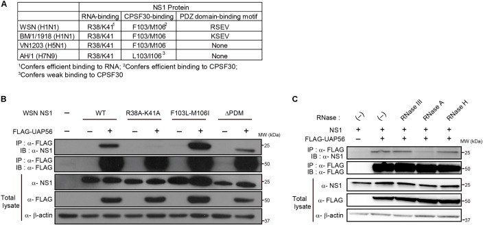
FIGURE 3.
Identification of NS1 residues critical for the UAP56–NS1 interaction. (A) Comparison of RNA-binding residues, CPSF30-interacting residues, and PDZ domain binding motifs among the NS1 proteins of WSN, BM/1/1918, VN1203, and AH1. (B) Interaction of UAP with mutant NS1 proteins. HEK293T cells were transfected with a protein expression vector for wild-type (WT) or mutant WSN-NS1 protein and FLAG-tagged UAP56 or a control vector. At 48 h post-transfection, the cells were lyzed and immunoprecipitated with anti-FLAG M2 antibody-conjugated magnetic beads. Co-precipitated proteins were analyzed by immunoblotting with anti-NS1 antibody. ΔPDM: deletion of the “PDZ domain-binding motif.” (C) Effect of RNase treatment on the UAP56–NS1 interaction. HEK293T cells were transfected with plasmids for the expression of WSN-NS1 and FLAG-tagged UAP56. At 48 h post-transfection, the cells were lyzed and the collected cell lysate was mock-treated or treated with the indicated RNases at 37°C for 20 min. The lysates were incubated with anti-FLAG antibody-conjugated magnetic beads, and co-precipitated proteins were analyzed by immunoblotting.

