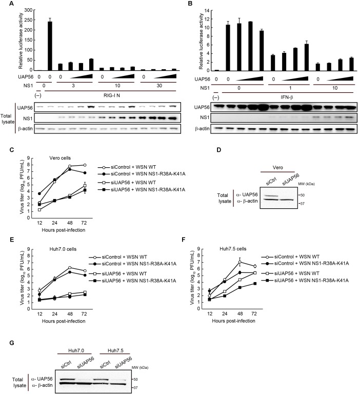
FIGURE 5.
Effect of UAP56 overexpression on the IFN-antagonist activity of NS1. (A) UAP56 overexpression does not affect NS1’s ability to suppress IFN-β promoter activity. HEK293T cells were transfected with plasmids expressing firefly luciferase under the control of an IFN-β promoter, pRL-TK luciferase (as an internal control), a constitutively active form of RIG-I (pCAGGS-RIG-I N), increasing amounts of pCAGGS-NS1 (0, 3, 10, or 30 ng), and pCAGGS-UAP56 (0, 3, 10, or 30 ng). Twenty-four hours later, the cells were lyzed and firefly and Renilla luciferase activities were measured. The firefly luciferase activity was normalized by the internal control value. The total amount of transfected vector was adjusted in all wells using the pCAGGS control vector. Data are shown as the mean ± SD (n = 3; biological replicates). UAP56 and NS1 expression levels in cells were examined by immunoblotting. (B) UAP56 overexpression does not affect NS1’s ability to suppress ISRE-driven gene expression. HEK293T cells were transfected with plasmids expressing firefly luciferase under the control of an ISRE promoter element, pRL-TK luciferase (as an internal control), and increasing amounts of pCAGGS-NS1 (0, 1, or 10 ng) and pCAGGS-UAP56 (0, 3, 10, or 30 ng). Twenty-four hours later, the cells were mock-treated or treated with IFN-β (10,000 U/mL), and cultured for 24 h. Luciferase activities were measured and data were analyzed as described in A. Data are shown as the mean ± SD (n = 3; biological replicates). UAP56 and NS1 expression levels in cells were examined by immunoblotting. (C–G) WSN virus or WSN-NS1-R38A-K41A mutant virus replication in cells transfected with siRNA targeting UAP56 or with a control siRNA. Vero (C), Huh7.0 (E), or Huh7.5 (F) cells were transfected with siRNA targeting UAP56, or a control siRNA. At 24 (Vero cells) or 48 h (Huh7.0 and Huh7.5 cells) post-transfection, the cells were infected with WSN virus or WSN-NS1-R38A-K41A mutant virus at an MOI of 0.01 and incubated at 37°C. Culture supernatants were collected at the indicated times post-infection and viral titers were analyzed by performing plaque assays in MDCK cells. The data are shown as the mean ± SD (n = 3; biological replicates). Levels of UAP56 expression in Vero (D) or Huh7.0 and Huh7.5 (G) cells transfected with siRNA targeting UAP56, or with a control siRNA. UAP56 expression levels were analyzed by immunoblotting at 48 (Vero cells) or 72 h (Huh7.0 and Huh7.5 cells) after siRNA transfection.

