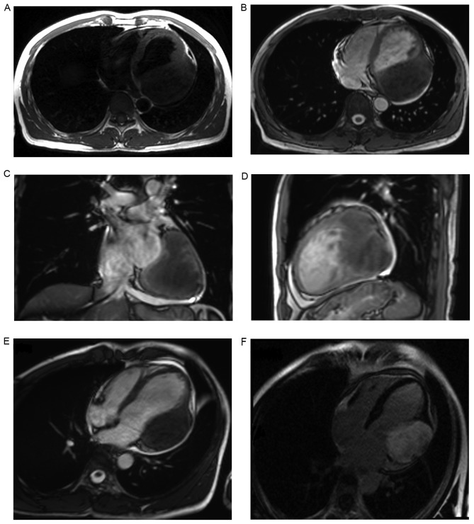
Figure 1.
Cardiac fibroma in a 43-year-old male patient. Magnetic resonance imaging indicated a large mass located in the lateral wall of the left ventricle of the heart. (A) T1 transaxial section. (B) T2 transaxial section. (C) T2 coronal section. (D) T2 sagittal section. The mass was T1 iso-intense and T2 hypo-intense relative to muscle. (E) Myocardial first-pass perfusion image indicated no enhancement in the tumor. (F) Delayed-phase perfusion image showed heterogeneous enhancement in the tumor.

