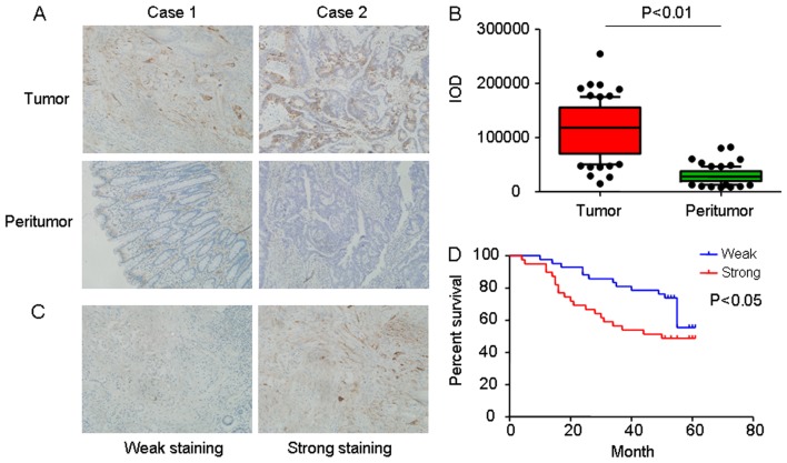
Figure 1.
Expression of Hsp27 in CRC tissues. (A) Representative immunohistochemical images of Hsp27 staining in matched tumor tissues and adjacent normal colorectal mucosal tissue (magnification, ×400). (B) A box and whisker plot (whiskers: 10–90%) of IOD for Hsp27 was obtained from TMA. (C) Representative expression levels of Hsp27 in CRC TMA, analyzed using immunochemistry analysis (weak staining, score + and negative; strong staining, score ++ and +++. Magnification, ×400). (D) Kaplan-Meier analysis of overall survival in 81 patients with CRC based on Hsp27 expression. Hsp27, heat shock protein 27; CRC, colorectal carcinoma; IOD, integrated optical density; TMA, tumor microarray.

