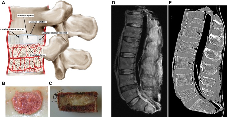
Fig 1.
(A) schematic drawing representative of functional spine unit and regions of the disc showing the caudal and cranial cartilaginous endplates, the nucleus pulposus and the anterior and posterior annulus fibrosus. The figure also shows the blood supply to the disc (B) Superior view of a spine unit after the removal of the posterior arch. (C) Sagittal view of the mid part of the spine unit, note the nylon stitch in the antero-superior region to maintain the fragment in functional position in all steps of the study. (D) MRI and (E) CT sagittal view with ligaments and paraspinal muscles intact for better contrast.

