Figure 3.
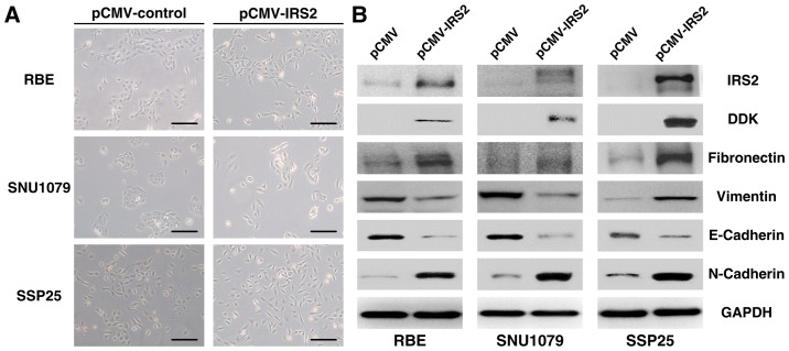
Effect of pCMV-IRS2 transfection on the EMT in RBE, SNU1079, and SSP25 cells. (A) After transfection with pCMV-IRS2 vectors, morphological changes from a cobblestone-like (left) to a spindle-like fibroblastic (right) appearance (magnification, ×50; scale bar, 100 µm). (B) RBE, SNU1079, and SSP25 cell extracts were subjected to 10% SDS-PAGE and western blot analysis with the respective primary antibodies against IRS2, vimentin, N-cadherin, fibronectin, and E-cadherin. GAPDH was used as an internal control. EMT, epithelial-mesenchymal transition; IRS2, insulin receptor substrate 2.

