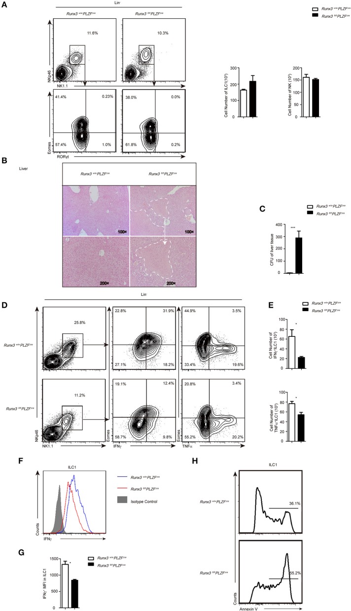
Figure 2.
Increased sensitivity to L. monocytogenes infection partially due to defective ILC1 function after Runx3 deletion. (A) Flow cytometry analysis of the percentage of ILC1s and NK cells in the liver from control and cKO mice before infection, and absolute cell number of the indicated ILC population. (B–H) Control and cKO mice were infected with L. monocytogenes through tail vein injection (n = 6 per group). (B) Hepatic histology of livers obtained from wild and cKO mice stained with haematoxylin and eosin. (C) Titres of L. monocytogenes in the liver were measured 48 h after infection. (D–G) Cells isolated from the livers of infected wild type or cKO mice were stimulated with PMA/ionomycin and BFA for 4 h. (D) Flow cytometry assay of intracellular IFNγ from ILC1s in the middle and intracellular TNFα on the right. (E) Absolute cell number of the indicated cell types from wild type or cKO mice in the liver. (F) The intracellular expression of IFNγ in ILC1s. Isotype controls are shaded curves, wild type groups are blue curves and cKO groups are red curves. (G) Mean fluorescence intensity (MFI) of IFNγ in ILC1s. (H) Apoptosis of liver ILC1s labeled with annexin V (n = 3) (mean ± SD of three samples in b, d and f; *P < 0.05; **P < 0.01 by Student's t-test). Data are from one experiment representative of five independent experiments with similar results in (A–F), and two independent experiments with similar results in (G).

