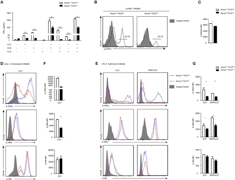
Figure 5.
The IL12R signaling pathway was defective after Runx3 deletion in ILC1s and NCR+ILC3s. (A) ELISA for IFNγ secretion by intestinal ILC1s from wild type and Runx3 KO mice after stimulation with the indicated cytokines labeled under each bar for 36 h. (B) Flow cytometry assay of p-STAT4 in intestinal ILC1s from wild type and Runx3 KO mice after stimulation with IL12. (C) Statistical analysis of the change of p-STAT4. (D,F) Wild type and Runx3 KO mice were infected with L. monocytogenes through tail vein injection (n = 3 per group). (D) The expression of IL12Rβ2, IL18Rα and IL15R in ILC1s from the liver after L. monocytogenes infection and (F) mean fluorescence intensity (MFI) of the indicated proteins in ILC1s. (E,G) Control and Runx3 KO mice were orally infected with S. typhimurium (n = 6 per group). (E) The expression of IL12Rβ2, IL18Rα, and IL15R on ILC1s and NCR+ILC3s from the small intestine after S. typhimurium infection and (G) mean fluorescence intensity (MFI) of the indicated proteins in ILC1s and NCR+ILC3s (mean ± SD of three samples in (A,C,F,G); *P < 0.05, **P < 0.01 and ***P < 0.001 by Student's t-test). Data are from one experiment representative of three independent experiments with similar results in (A–C) and four independent experiments with similar results in (D–G).

