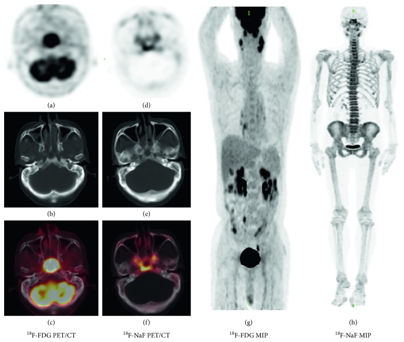
Figure 1.
A 63-year-old man was diagnosed with nonkeratinizing nasopharyngeal carcinoma. (A–C) transverse sections of PET, CT, and fusion views in 18F-FDG PET/CT, respectively. (D–F) transverse sections of PET, CT, and fusion views in 18F-NaF PET/CT. (G and H) the maximum intensity projection (MIP) of 18F-FDG PET/CT and 18F-NaF PET/CT, respectively. Skull-base invasion was revealed on 18F-NaF PET/CT but was hidden on 18F-FDG PET/CT because of the interference from the tumor tissue. This was consistent with MRI two days before 18F-NaF PET/CT.

