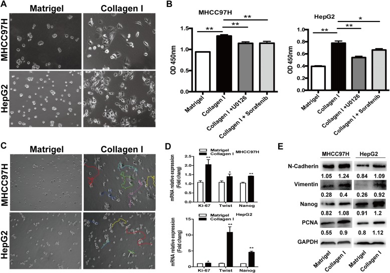
Fig. 2.
Collagen I stimulated the proliferation, motility, and the expression of EMT and progenitor-like markers in heat-treated residual HCC cells. a Compared with the cells cultured on Matrigel, heat-treated residual HCC cells on collagen I displayed a proliferative, protrusive and spindle-like appearance. b Collagen I promoted proliferation of heat-treated residual HCC cells as determined by the WST-1 proliferation assay. The OD (optical density) was measured at 450 nm wavelength. c Compared with Matrigel, collagen I enhanced the motility of heated-exposed residual HCC cells as demonstrated by tracking analysis. d As shown by qRT-PCR, the mRNA expression of Ki-67, twist, and Nanog was increased in heat-exposed residual HCC cells cultured on collagen I versus Matrigel. e The increased expression of PCNA, vimentin, N-cadherin and Nanog protein in heat-exposed residual HCC cell cultured on collagen I was detected by Western blot. Expression levels of target proteins were normalized to the corresponding levels of GAPDH. **, P < 0.01; *, P < 0.05

