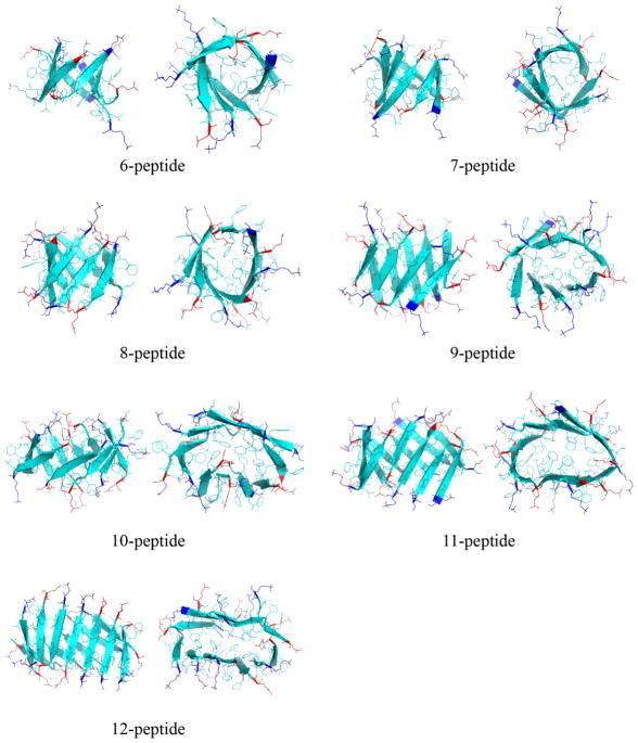
Figure 9.
Representative structures of β-barrels with various sizes are shown in both side and top views. Each peptide is shown in cartoon and all residues are shown as sticks. Positively-charged lysine residues in the N-terminal are colored blue and negatively-charged glutamate residues in the C-terminal are colored red, while the rest hydrophobic residues are colored cyan.

