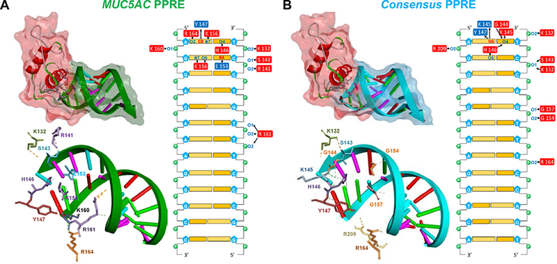
FIGURE 7. The PPARγ-DBD interacts with nucleotides in the MUC5AC PPRE1.

Nucleic acid-protein docking was performed as described in Materials and Methods. Cartoon representation and stereo views of the predicted PPARγ-DBD in complex with (A) MUC5AC PPRE1 and (B) the consensus PPRE. The PPARγ-DBD is depicted in secondary structure with a red surface. The amino acids in contact with the DNA are depicted as colored sticks. The DNA backbone is represented as green (the MUC5AC PPRE1) or sky blue (the consensus PPRE) arrows with semi-transparent surfaces of the same color while nucleotides are drawn as base-colored sticks. Hydrogen bonds are indicated with green dashed lines. Contact maps illustrate interacting amino acids within a 4.0 Å distance from the nucleotides. Donors and acceptors are colored in red and blue, respectively. Arrows indicate hydrogen bonding.
