
FIGURE 8. PPARγ plays a key role in regulating mediators of inflammation and mucus production in human bronchial epithelial cells.
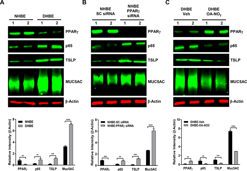
NHBE and DHBE cells were cultured as indicated in Materials and Methods. PPARγ was silenced with siRNA in NHBE cells or activated by OA-NO2 in DHBE cells. Western blotting was performed to determine the levels of indicated proteins (PPARγ, NF-κB/p65, TSLP, and MUC5AC) in (A) untreated NHBE and DHBE cells, (B) NHBE cells treated with scrambled (SC) siRNA or PPARγ-specific siRNA, or (C) DHBE cells treated with vehicle (Veh) or 1 μM OA-NO2 for 6 h. β-Actin served as a loading control. The results were reproduced at least two times independently. Image quantification was performed with MATLAB Image Processing Toolbox. The data are expressed as the mean ± SD; **P < 0.01, ***P < 0.001.