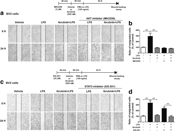
Fig. 8.
Ibrutinib alters LPS-induced BV2 microglial cell migration through AKT signaling, but not STAT3 signaling. a BV2 microglial cell monolayers were scratched with a fine tip, pretreated with MK2206 (AKT inhibitor, 3 μM) or vehicle (1% DMSO) for 30 min, treated with ibrutinib (500 nM) or vehicle (1% DMSO) for 30 min, and then treated with LPS (100 ng/ml) or PBS for 23 h. Images of the wound gap were acquired at 0 h (i.e., immediately after scratching) and after 24 h. b Quantification of data from a (con, n = 41; LPS, n = 42; ibrutinib+LPS, n = 41; MK2206+LPS, n = 38; ibrutinib+MK2206+LPS, n = 41). c BV2 microglial cell monolayers were scratched with a fine tip, pretreated with S31-201 (STAT3 inhibitor, 25 μM) or vehicle (1% DMSO) for 30 min, treated with ibrutinib (500 nM) or vehicle (1% DMSO) for 30 min, and then treated with LPS (100 ng/ml) or PBS for 23 h. Images of the wound gap were acquired at 0 h (i.e., immediately after scratching) and after 24 h. d Quantification of data from c (con, n = 24; LPS, n = 25; ibrutinib+LPS, n = 26; S31-201+LPS, n = 25; ibrutinib+S31-201+LPS, n = 24)

