Figure 2.

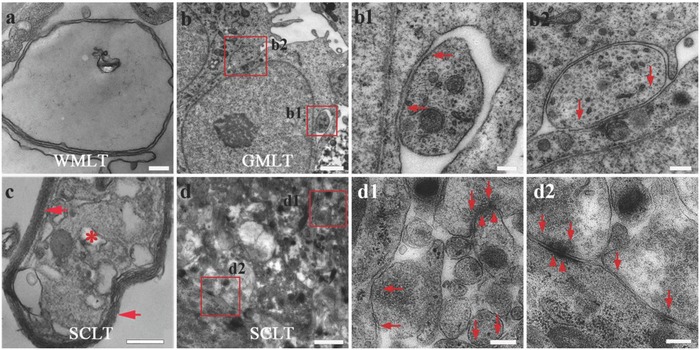
Myelin and synaptic formation potential of the SCLT. a,b,b1,b2) The singly cultured WMLT or GMLT module presents features of immature myelin sheath (a), or synaptic connections (arrows indicate small and dense vesicles in (b), (b1), (b2)) under the electron microscope, respectively. c) The SCLT shows multilamellar myelin sheaths (arrows in (c)) enwrapping an axonal profile (asterisk in (c)) with (d), (d1), (d2) seemingly more mature features of synapses, like the presynaptic vesicles (arrows in (d1), (d2)) and the postsynaptic membrane density (arrowheads in (d1), (d2)). Scale bars = 200 nm in panels (a), (b1), (b2), (c), (d1), and (d2), 1 µm in panels (b) and (d).
