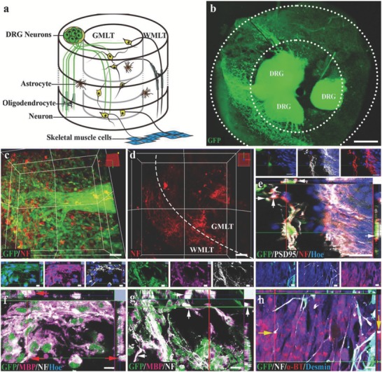
Figure 4.

The SCLT establishes connections with DRG or muscle cells. a) A schematic diagram showing a SCLT establishes connections with the organotypic cultured DRG or muscle cells. b) GFP donor–derived DRG extended NF positive neurites into the c,d) SCLT. e) The neurons in GFP donor–derived GMLT region form close contacts with DRG (from GFP negative donor), with PSD95 expression at the contact sites. f,g) GFP donor derived WMLT of a SCLT‐expressed MBP (arrows) wrapping NF‐positive DRG neurites (GFP negative in (f)) or neurites (GFP negative in (g)) extending from the neurons in the GMLT region. h) The outgrowing NF positive neurites from GFP positive SCLT form close contacts with α‐BT‐expressing muscle cells with bouton‐like enlargement (arrows) at the terminal. Scale bars = 500 µm in panel (b), 50 µm in panels (c) and (d), 20 µm in panels (e)–(h).
