Figure 6.

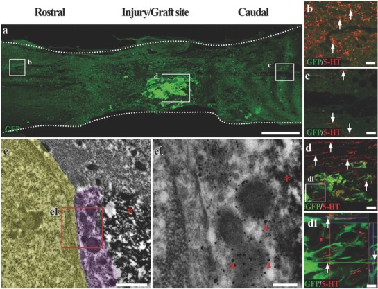
The donor neurons in the SCLT integrate with descending 5‐HT nerve fibers. a) A longitudinal section of spinal cord containing GFP donor derived GMLT of a SCLT. b,c) 5‐HT positive descending nerve fibers passed through the injury/graft site of spinal cord and extended several millimeters into the caudal area to the injury/graft site 8 weeks after SCLT implantation (c). d,d1) Most 5‐HT nerve fibers traveled longitudinally through the WMLT region, while some of them formed close contacts with the donor cells in the GMLT (arrows). e,e1) IEM showed that a 5‐HT nerve fiber (superimposed in light purple and labeled by nanogold particles, arrowheads in (e1)) formed close connection with GFP positive donor cells (DAB labeled, asterisks) and the host cell (superimposed in yellow). Scale bars = 500 µm in panel (a), 40 µm in panels (b)–(d), 20 µm in panel (d1), 1 µm in panel (e), 200 nm in panel (e1).
