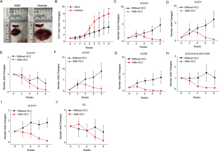
Figure 1.
Establishment of patient-derived xenograft (PDX)-hepatocellular carcinoma (HCC) humice model and the blood immune cell number changes. (A–B) PDX tumours were transplanted subcutaneously to NOD-scid Il2rg−/− (NSG) mice and humice (n=5) aged 8–10 weeks. (A) Representative image of tumours and spleens 8 weeks after transplantation in NSG and humice. (B) The weekly changes in PDX tumour size in NSG and humice after transplantation. Data are presented as fold changes normalised to the size of tumour before PDX transplantation (week 0). *P<0.05, **P<0.01. (C–J) PDX tumours were transplanted subcutaneously to humice aged 8–10 weeks. Blood immune cell frequencies and absolute numbers from humice without tumour (n=5) and humice with tumour (n=5) were analysed biweekly by flow cytometry. Data are presented as fold changes normalised to the cell numbers of specific cell types before PDX transplantation (week 0): human CD45+ (hCD45+) (C), hCD3+ (D), hCD19+ (E), hCD4+ (F), hCD8+ (G), hCD14-HLA-DR-CD56+ (H), hCD14+ (I) and DC (J).

