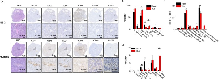
Figure 4.
Infiltration of human immune cells in tumour. Tumours were harvested at 8 weeks postengraftment and analysed for human immune cell infiltration (n=5). (A) In situ stain of various human immune cell types infiltrated in hepatocellular carcinoma (HCC) tumours from NOD-scid Il2rg−/− (NSG) and humice. (B) The frequencies of major human immune cells from blood and HCC-patient-derived xenograft (PDX) tumour analysed by flow cytometry. (C) The proportions of T cell subtypes within T helper and cytotoxic T cells from blood and HCC-PDX tumour. (D) The proportions of myeloid subsets from blood and HCC-PDX tumour. *P<0.05, **P<0.01. TIL, tumour-infiltrating leucocytes.

