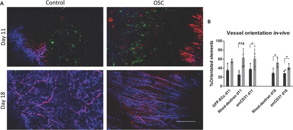
Figure 5.

Implantation of oscillatory stretched vascularized grafts into a mouse dorsal window chamber. A) Confocal images of the penetrating host vessels into the implanted oscillatory stretched and static control constructs cultured for 21 d in vitro, stained with rhodamine‐dextran (red) and anti‐mouse CD31 (blue) that were injected via the mouse tail vein. Scale bar = 500 µm. B) Quantification of vessel orientation; *p < 0.05, **p < 0.01, ***p < 0.001.
