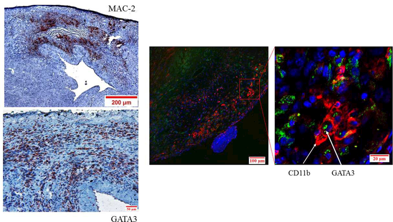
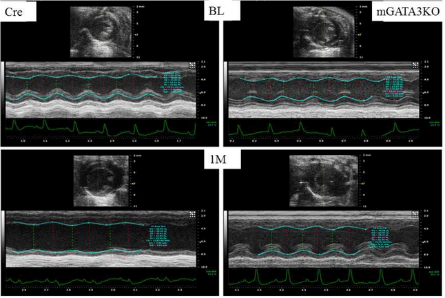
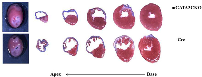
Figure 1. Deficiency of GATA3-Positive Macrophages Improved Cardiac Function After MI.
(A) Representative immunostaining of LV sections from control Cre mice. Serial sections of LVs from control Cre mice at 8 days post-MI were stained with anti–Mac-2 or anti-GATA3 antibodies. GATA3-positive staining is clearly visible in the infarcted region of the myocardium, which contains large numbers of macrophages, but not in the healthy region. Sections were also stained with fluorescent-labeled antibodies to demonstrate the nuclear colocalization of GATA3 in macrophages located in the infarcted region of the myocardium. (B) Representative M-mode echocardiograms from the 2 groups of mice before (BL) and 1 month after MI (1M). (C) Representative photographs of Masson Trichrome staining from sections from the base to the apex of hearts from the 2 mice genotype groups. Photographs of all of the hearts are shown in Online Figure 3. (D) Morphometric analysis of each heart section. The number of animals in each group is shown in Table 1. LV = left ventricle; mGATA3KO = myeloid-specific GATA3 knockout mouse; MI = myocardial infarction.


