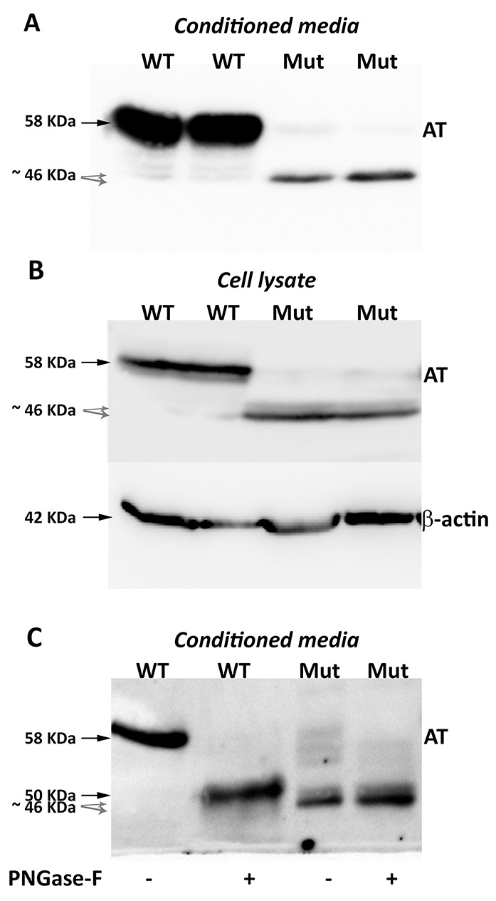
Figure 2. Antithrombins expressed by HEK-EBNA cells 24 h after transfection with pCEP4-S169A (WT) and pCEP4-S169A-M1I (Mut) plasmids.

Proteins were identified by SDS-PAGE and western blot using an anti-human antithrombin polyclonal antibody. (A) Antithrombin (AT) released to the conditioned media. The same volume of conditioned was used for cells transfected with both plasmids. (B) Intracellular antithrombin and beta-actin expression. The same amount of cell lysates was used for cells transfected with both plasmids. (C) Glycomic analysis of recombinant antithrombins purified from the conditioned media. The wild-type protein and the small antithrombins were treated (+) or not (-) with N-glycosidase F (PNGase-F) and detected by western blot. Black arrows point the wild-type antithrombin. Grey dashed arrows indicate the small variants. Estimated molecular weights are also indicated.
