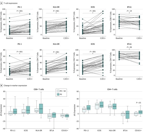
Figure 2. Effect of Ipilimumab Administration on Peripheral T-Cell Phenotype.
Best response was assessed by Response Evaluation Criteria in Solid Tumors, 1.1 for the 34 evaluable patients; 8 patients were not evaluable because they were removed from study therapy before the first protocol-mandated computed tomography scan at week 12. All patients were assessed for toxic effects. Statistical analysis of the changes in the expression of immune markers was assessed in correlation with clinical response. Both CD4+ and CD8+ T cells demonstrated significantly increased expression of programmed cell death protein 1 (PD-1), human leukocyte antigen–antigen D related (HLA-DR), and inducible T-cell costimulator (ICOS) following treatment compared with pretreatment baseline. A, With ipilimumab treatment, CD4+ and CD8+ T cells showed an increase in expression of PD-1, HLA-DR, and ICOS but not B- and T lymphocyte–associated markers after the therapy. B, Changes in the expression of CD8 and CD4 markers were not associated with clinical response. Change in the CD161+ expression in CD4+ T cells, however, was significantly higher in patients whose disease progressed compared with those who had stable disease (SD) or partial response (PR). The horizontal line in each box indicates the median, and the top border of the box indicates the 75th percentile, the bottom the 25th. The whiskers above and below the box mark represent minimum and maximum values for that particular graph. BTLA indicates B- and T-lymphocyte attenuator; PD, progressive disease.

