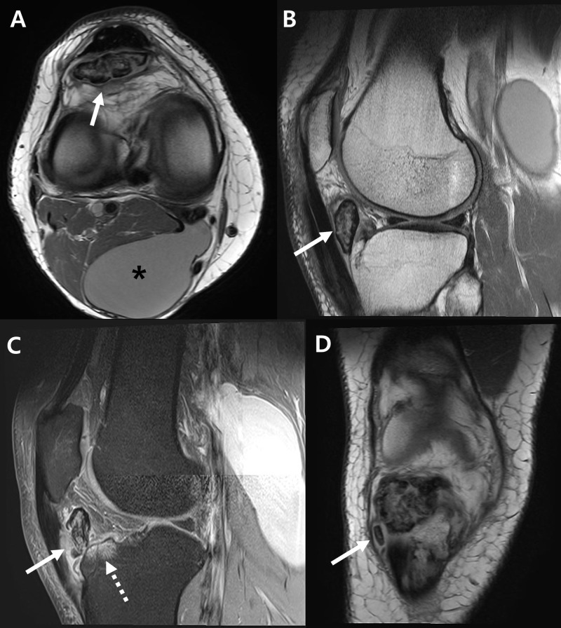
Figure 2. Right knee magnetic resonance images.
(A) A large ossicle with heterogenous signal intensity (arrow) and a Baker’s cyst (asterisk) are seen on axial image. (B) From T1-weighted sagittal image, the ossicle (arrow) is separately located inside the infrapatellar fat pad. (C) T2-weighted sagittal image shows inflammation of patellar tendon (solid arrow) and bone marrow edematous change of anterior tibia plateau (dotted arrow). (D) Small separated ossicle (arrow) was found inferior to the large ossicle from coronal image.

