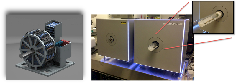
Fig. 1.

Left: A rendering of the PET scanner design. 45 PET detectors are arranged in 5 rings to provide a scanner diameter of 7.6 cm & axial length of 13 cm, making it suitable to image both whole-body rat & mice. Right: The MOLECUBES β-CUBE PET (right) and X-CUBE CT (left) scanners installed in the Small Animal Imaging Facility at the University of Pennsylvania. Both PET and CT scanners are compact and have a very small footprint to enable bench-top imaging. Inset on the right shows the animal bed retracted from the PET scanner. It can be easily detached and translated between the PET & CT scanners, obtaining automatically co-registered 3D PET & CT images.
