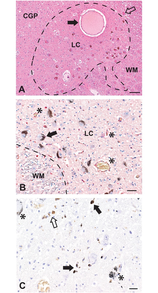
Fig 1. Normal appearance and pathology of the locus ceruleus.
(A) The locus ceruleus (LC, outlined) contains numerous, mostly neuromelanin-containing, neuronal cell bodies. White matter (WM) is present at the right, and the central grey of the pons (CGP) at the left. A large thin-walled blood vessel (closed arrow), probably a post-capillary venule, is seen within the locus ceruleus. A mesencephalic trigeminal neuron is seen in the upper right corner (open arrow). Hematoxylin and eosin, Bar = 100 μm. (B) The capillary density in the locus ceruleus is slightly increased, compared to adjacent white matter (WM), as seen by the red-immunostained capillary endothelial cells (some asterisked). Black AMG grains are present in several locus ceruleus neurons (one with arrow). AMG/hematoxylin/CD31, Bar = 50 μm. (C) This locus ceruleus has a reduced density of neurons. Free and macrophage-bound neuromelanin originating from destroyed locus ceruleus neurons is prominent, with this pigment having either AMG (closed arrow) or no AMG (open arrow). Some remaining locus ceruleus neurons either with (asterisks) or without AMG are seen. AMG/hematoxylin, Bar = 20 μm.

