Figure 2.

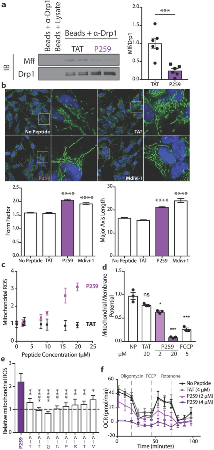
P259 inhibits the Mff/Drp1 protein-protein interaction and physiological mitochondrial fission in SH-SY5Y cells, resulting in mitochondrial dysfunction. (a) Co-immunoprecipiation of Drp1 and Mff from SH-SY5Y cell lysate treated with 1 µM P259 for 1 hour (n = 3, with 2 replicates). Represented Western blot is cropped from a full blot shown in the Expanded Western Blots section of the Supplementary Information. (b) Immunofluorescence images depicting mitochondrial morphology after a 30-minute treatment with 4 µM TAT, 4 µM TAT-P259 (P259), or 10 µM Mdivi-1. Hoechst 33342 is shown in blue and Tom20 in green (n = 3, 10 images each). Below, quantification of the form factor (degree of branching) and major axis length (length of mitochondria). At least 6,000 mitochondrial fragments were measured for each condition. Mitochondrial reactive oxygen species (ROS) production (MitoSox Red) (c) and mitochondrial membrane potential (TMRM) (d) after overnight treatment with indicated concentrations of peptide or FCCP. Data are from 3 independent experiments per condition normalized to the average no peptide value. (e) Relative mitochondrial ROS (MitoSox Red) signal in GFP positive cells expressing full-length or alanine-substituted P259 at indicated amino acids after two days of transfection. Values are normalized to a GFP empty vector (n = 3, with 3 replicates). (f) The effect of overnight treatment with 4 µM TAT or P259 on electron transport chain flux as measured through the oxygen consumption rate (OCR) (n = 3, with at least 3 replicates). Data are mean ± s.e.
